RATOC
       RATOC >BONE>Bone mineral density measurement
            Analysis service    Bone    Tissue cell reconstruction    Fiber    Otolith    Display options       Particle measurement    Through-hole measurement    4Dkinetic Anlysis      
            Cardiac function measurement        Light scattering tomograph        User support


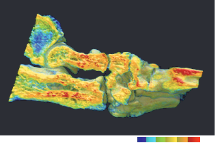
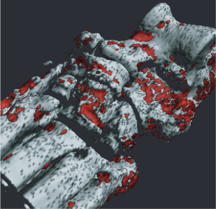
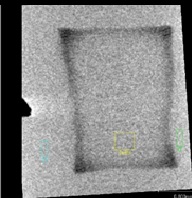
3D Bone BMD measurement
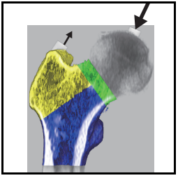
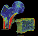
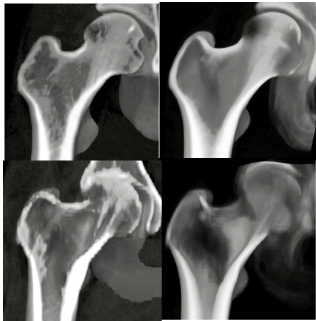
          

Femur measurement
          

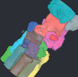
Waist, chest, cervical spine
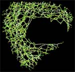
          

bone morphometry
          

Bone mineral density measurement
          

Rheumatism measurement
          

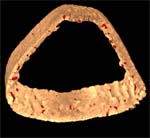
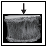
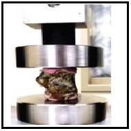
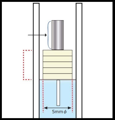
Cortical bone measurement
          

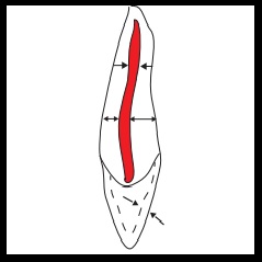
Tooth 3D morphological detail measurement
          

Medication measurement software using the VBMmethod
        

Measurement of enamel mineral loss
          

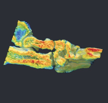
Finite element analysis
          

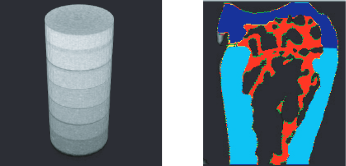
Bone Mineral Phantom
          
Bone mineral density measurement      TRI/3D-BON-BMD
Using high-resolution ƒÊCT images, bone marrow, cancellous bone, and cortical bone
can be separated.
BMD value of true cancellous bone and cortical bone without bone marrow
can be obtained.
3D display of BMD value in pseudo color.
You can see the distribution of bone density for each part at a glance.

1. Calibration curve creation

CT value and BMD value using a phantom
with known BMD Create standard curve
Phantom 3D image (image)

           Separation of cancellous bone,
cortical bone and bone marrow

2DExtraction of BMD bone mineral content measurement sites

3DMeasurement item

@ Measure BMD value and bone mineral content for each part
 AverageCT                  : average CT value
  Bone density [mg/c]       : mg/cm3 on the label
  Volume [cm3]              : Total volume of label [cm3]
  Bone mineral content [mg] : Labeled bone mineral content [mg]
  Bone Density Min          : Minimum bone density of the label
  Bone Density Max          : Maximum bone density of the label
  Standard deviation        : standard deviation of bone density