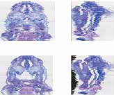
3D reconstruction software(SRFIII)
virtual slide(SRF-VS)
Pathological tissue 3D reconstruction/
observation software
Large-scale image analysis
|
3D reconstruction software(SRFIII) |
virtual slide(SRF-VS) |
Pathological tissue 3D reconstruction/ observation software |
Large-scale image analysis |

|
|
feature |
|
1. Manual tracing is almost never required. Automatically extract contours from color images. 2. The 3D construction image can be rotated and cut in real time, and a movie can be displayed. 3. Saved data in TRI/SRF can be displayed. |
| Measurement DaTa |
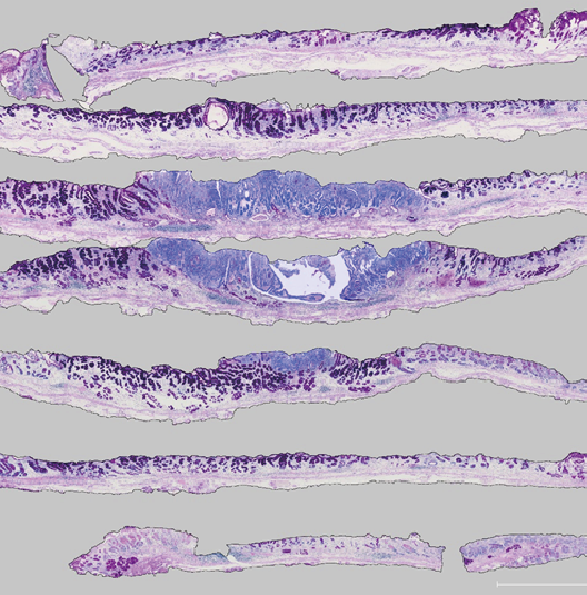
Print out the microscope serial section images and trace the outline of the tissue
with a colored felt-tip pen. It is read by the scanner and used as an input image
to SRF-R.
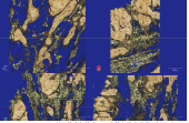
Input images to SRF-R: Print out serial section images,
The outline of the tissue is traced with a red felt-tip pen 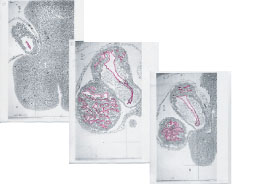 Mouse embryo gestation day 11 part of serial section (Epon embedded, 1 μm thick) Saitama Medical University Anatomy 2nd Professor Tamiko Hiruma Provided 29 sheets 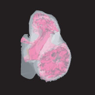 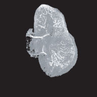 Image generated from extracted contours Volume image of outline |
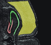
| 3D construction example in SRF |
 Cerebellar nucleus-derived nerve endings and surrounding structures in feline thalamic VL nucleus Doc Futami Sato 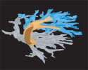 Fetal lung bronchus reconstructed from 50 light microscope section images 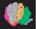 Rat kidney and glomeruli prepared from 30 serial electron microscope sections Mr. Nobuaki Yamanaka Ontoshi |
| From input to output |
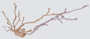
INPUT  Trace a photo with color pens scanner input 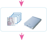 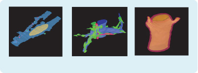 Rat intestinal/cell bile duct and surrounding vessels Coronary arteries with intimal thickening <Provide materials> Waseda University Akita University School of Medicine Kyushu University School of Medicine Professor Terumasa Komuro Professor Tetsuo Kato Hiroaki Yoshino Professor Yutaka Nakajima |