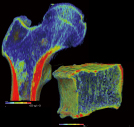
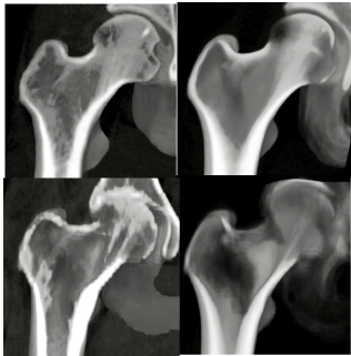
3D Bone BMD measurement
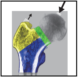
Femur measurement
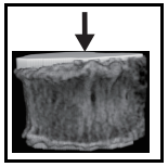
Waist, chest, cervical spine
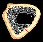
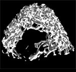
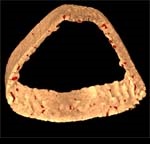
bone morphometry
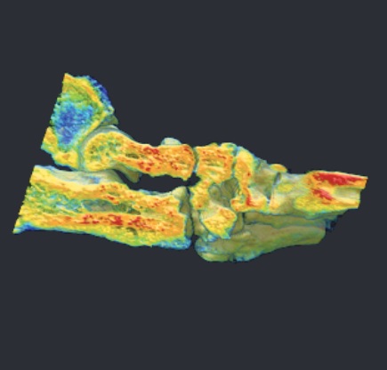
Bone mineral density measurement
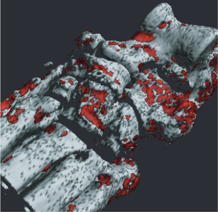
Rheumatism measurement
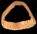
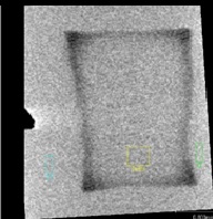
Cortical bone measurement
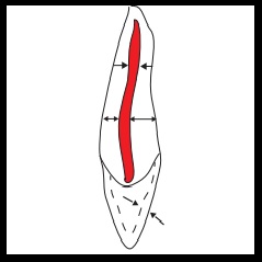
Tooth 3D morphological detail measurement
Medication measurement software using the VBMmethod
Measurement of enamel mineral loss
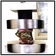
Finite element analysis
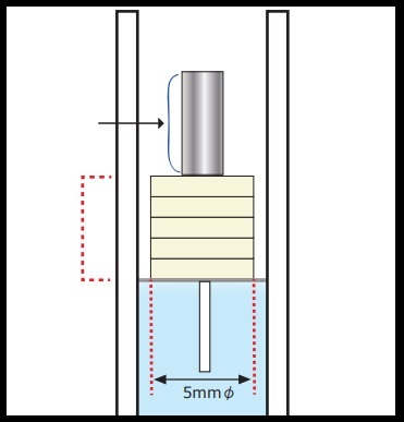
Bone Mineral Phantom

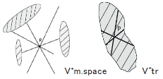
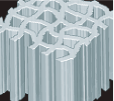
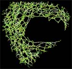
 TBPf < 0 TBPf ≒ 0 TBPf > 0
SMI ≒ 0 SMI ≒ 0 SMI ≒ 3.0
The medullary cavity The cancellous bone extends It grows like a bar.
is partitioned in a in a plate-like shape.
honeycomb pattern The medullary cavity is not
stretched like a bar divided into compartments.
by platy cancellous
bones. surrounded by
concave walls of
small chambers
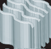
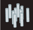
TBPf < 0 TBPf ≒ 0 TBPf > 0
SMI ≒ 0 SMI ≒ 0 SMI ≒ 3.0
The medullary cavity The cancellous bone extends It grows like a bar.
is partitioned in a in a plate-like shape.
honeycomb pattern The medullary cavity is not
stretched like a bar divided into compartments.
by platy cancellous
bones. surrounded by
concave walls of
small chambers