Virtual slide (VS) image 3D reconstruction TRI/3D-SRF-VS
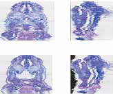
Input image
Virtual slide image format Tif, jpg, raw file, etc.
scanner input
Can handle multiple sections arranged on a slide
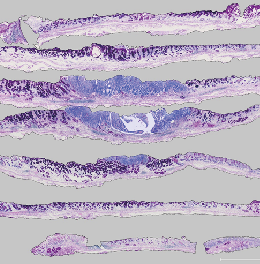
Automatic alignment of section images
Aligns the misalignment between serial intercepts in the input.
Method
automatic alignment
A specific tissue is labeled to minimize the positional deviation
between figures with the same label.
Feature point registration
It is effective when there are target alignment markings in the specimen or
when there is a large tissue with small positional deformation.
manual alignment
scroll, rotate
tissue extraction
Tomographic cutout
You can decide the observation direction and the measurement direction of the tissue.
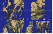
color extraction
Extract specified color from RGB color image
Binarization, 3D binary image processing
Binarization and binary image processing for tissue extraction
are performed simultaneously on continuous tomograms.
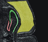
interpolation extraction
If automatic extraction is difficult, the contour is traced at a rate of 1 sheet for multiple sheets,
and the section in between can be obtained by contour interpolation.
Sectioning of extracted tissue
Only the extracted tissue is cut out from the original image and displayed.
The 3D shape of only the target tissue can be observed without being blocked by other tissues.