RATOC
       RATOC >tissue cell reconstruction>large scale analysis
            Analysis service    Bone    Tissue cell reconstruction    Fiber    Otolith    Display options       Particle measurement    Through-hole measurement    4Dkinetic Anlysis
            Cardiac function measurement        Light scattering tomograph         User support


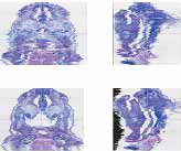
3D reconstruction software(SRFIII)


virtual slide(SRF-VS)


Pathological tissue 3D reconstruction/
observation software

Large-scale image analysis          

Large-scale image analysis   TRI/FCS-NUC64

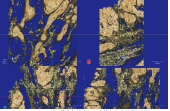
Extracting tissues and cells from various stained tomographic images
and reconstructing 3D images

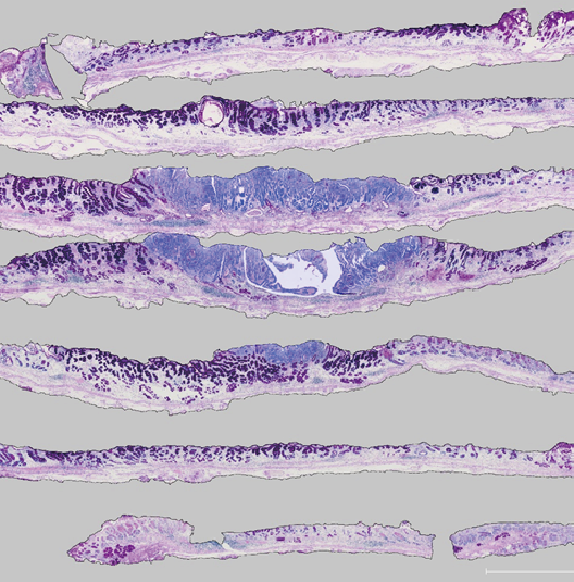
3D reconstruction of tiled laser scanning microscope images
and virtual slide images (VS).
Display analysis of 10,000 x 10,000 pixels x 5,000 tiling images is possible.

Feature point

  • Large data display
    A 3D image of a 5 mm x 5 mm x 5 mm tissue obtained by tiling a laser scanning
    microscope image with a resolution of 1 μm or more can be displayed continuously
    from the entire display to an enlarged image of 1 μm pixel.
  • Cell nucleus extraction batch processing
    Nucleus extraction operations performed from dozens of tomographic images can be
    batch processed in a night job for 5,000 images.
  • Brain tissue cell nucleus extraction
    Cell nuclei can be extracted from brain tissue images obtained by tiling.
  • Mouse brain tissue summary measurement
    It is possible to measure the number density of nuclei for each tissue site.

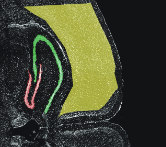
  •           LSMimage                     nuclear image                 Nucleus + LSM image

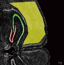
    Nuclear measurement ROI (region of interest)
  • red:  High density site
  • yellow: Low density site


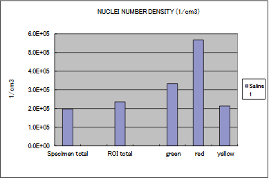
  • Nuclei contained within the whole sample
    ROItotal Nuclei included in all areas within ROI
    green green Nucleus in ROI
    red  redNucleus in ROI
    yellow yellowNucleus in ROI