RATOC
      RATOC > Tissue cell reconstruction>3D reconstruction software(SRFIII)
            Analysis service    Bone    Tissue cell reconstruction    Fiber    Otolith    Display options       Particle measurement    Through-hole measurement    4Dkinetic Anlysis
            Cardiac function measurement        Light scattering tomograph         User support


3D reconstruction software(SRFIII)


virtual slide(SRF-VS)


Pathological tissue 3D reconstruction/
observation software

Large-scale image analysis          

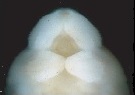
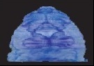
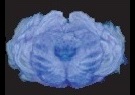
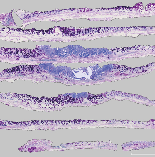
Tissue cell serial section 3D reconstruction software   TRI/3D-SRFIII

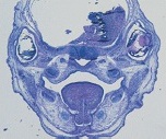
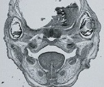
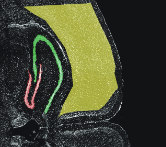
Extracting tissues and cells from various stained tomographic images
and reconstructing 3D images

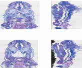
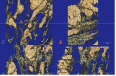
There are various staining methods depending on the tissue
example
 Shape stained with HE
 Calcified tissue stained by von Kossa
Alignment synthesis of these is possible

Victoria blue-HE


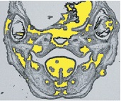
Color extraction cross-sectional image
Extract the stained cross-sectional image


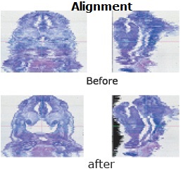
Automatic alignment of section images

  • Aligns the misalignment between serial intercepts in the input.
    Method
    • Alignment between feature points
      This is effective when there are alignment markings such as
      corners in the specimen,
      or when there is a large tissue with small positional deformation.
    • Label specific tissue and automatically align the center of gravity
      and direction of the same labeled figures
    • manual alignment
      zoom, scroll, rotate


      Macroscopic view and 3D constructs of an endothelin-1 (-/-) embryo (E16.0)