


Int. J. Plant Sci. 169(7):908-917. 2008.
Department of Enviromental Sciences, Faculty of Science, Niigata University, Japan
Department of Palaeobotany, Swedish Museum of Natural History
Japan Synchrotron Radiation Research Institute
Department of the Geophysical Sciences, University of Chicago
The following detail of movies and images were published on The University of Chicago
'Int. J. Plant Sci. 167(7):908-917.2008.'
|
|
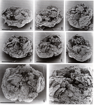 |
Fossil flower of Futabanthus asamigawaensis gen. et sp. nov. (Annonaceae) from the Ashizawa Formation (Futaba Group), Late Cretaceous (early Coniacian), of northeastern Honshuu, Japan, specimen PP 53685 ; SEM.A-F, Oblique lateral views of flower rotated counterclockwise to reveal the relative positions and shapes of perianth parts, androecium, and gynoecium. Scale bars = 1 mm. G, Flower from above, showing remains of two whorls of tepals (t), numerous stamens (s) comprising the androecium, and carpels comprising gynoecium (g). Scale bar = 1 mm. H, Oblique lateral view of center of flower, showing carpels on the central conical projection of receptacle surrounded by incurved stamens. Scale bar = 0.5 mm. | ||
|
|
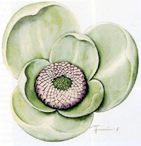 |
Reconstruction of Futabanthus asamigawaensis gen. et sp. nov showing two trimerous perianth cycles, numerous stamens, and numerous carpels. Flower illustrated with tepals opened to reveal the androecium and gynoecium. At anthesis, many flowers of extant Annonaceae are more tightly closed. | ||
|
|
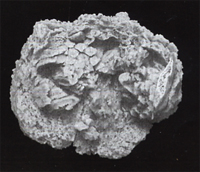 |
Image from a three-dimensional animation, based on micro CT scans, that shows overall floral structure. A video is available in the online edition of the International Journal of Plant Sciense. | ||
|
|
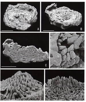 |
Fossil flower of Futabanthus asamigawaensis gen. et sp. nov. illustrated in fig. 1. A-C, E, F, Micro-CTimages. D, SEM. A, Flower from above, with stamens removed to reveal numerous carpels borne on the central projection of the receptacle. Scale bar = 1 mm. B, Oblique lateral view of flower with stamens removed, showing the closely packed carpels on the central projection of the receptacle. Scale bar = 1 mm. C, Section through flower perpendicular to plane of the receptacle disk, showing carpels in longitudinal section. Scale bar = 1 mm. D, Detail of distal parts of carpels. Scale bar = 0.1 mm. E, Lateral view of closely packed carpels revealed after stamens are removed from the micro-CT image. Scale bar = 0.5 mm. F, Longitudial section through carpels. Note larger cavities in the receptacle that may be the remains of oil cells. Scale bar = 0.5 mm. |
||
|
|
|
Images from a three-dimensional animation, based on micro CT scans, that shows serial sections parallel to the place of the flattened floral receptacle. A video is available in the online edition of the International Journal of Plant Sciences. |